Imagine walking down a dark pathway at night. You turn on a flashlight and suddenly you are alerted to things you couldn’t see before that could trip you up — tree roots, uneven ground, winding turns. That’s what a 3-D cone beam imaging system does inside a patient’s mouth.
It takes comprehensive, highly precise images that highlight the oral cavity and surrounding anatomy in ways that traditional 2-D X-rays, the standard of care, simply cannot.
Dr. Daniel Butterman is a general dentist in Arizona who invested in cone beam technology for one reason — to gain more confidence in diagnosis and implant treatment planning. However, he found that it was not just powerful but also adaptable to almost every type of dentistry he was doing, including endodontics work.
Cone beam applications in endodontics
Locating canals: Root canals are complex procedures, and one reason is the challenge of mapping the canals to be sure nothing is missed and every part of the infection is dealt with successfully. To have the best chance for a good outcome, all of the canals should be seen and become a part of the treatment plan.
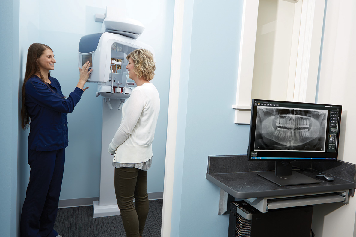
3-D imaging helps endodontists see how many canals there are and where they are located. (Photo: Henry Schein Dental)
3-D imaging helps endodontists see how many canals there are and where they are located. Doctors save a lot of time because they can see immediately if there are unusual anomalies, like a second mesiobuccal canal or missed lingual canals, or roots right on top of sinus cavities.
If there’s an anomaly, endodontists know before beginning the procedure and can adjust their treatment plan accordingly. In fact, a study evaluated how treatment plans were affected when cone beam imaging data was added to cases vs. only traditional X-rays.
It found in 62 percent of the cases, evaluators changed their treatment plans on the strength of the 3-D images. This proves it significantly influences the direction and effectiveness of a patient’s treatment, when made available, presumably for the better.
Discovering other unusual pathologies: 3-D imaging can also help endodontists see other conditions that are difficult, if not impossible, to see in intraoral X-rays: vertical root fracture, cracked teeth or interior or exterior root reabsorption. A clinical assessment, which asks a lot of questions looking for red flags, is done alongside the 3-D image.
Diagnosing pain: Most doctors have been frustrated when they are unable to find a reason for patient pain because X-rays and other clinical assessments haven’t located the source. Butterman uses his cone beam with almost every patient as he believes it gives him so much detailed information to discover the source of pain, helping solve problems that were previously mysteries.
“I can’t tell you how many patients have come in with massive infections in existing root canals,” said Butterman, who adds that previous 2-D X-rays showed no signs of trouble.
In a 2015 joint position statement of the American Association of Endodontists and the American Academy of Oral and Maxillofacial Radiology, cone beam imaging was recommended as the standard for patients with “contradictory or non-clinical signs” because the cone beam was able to detect problems earlier than traditional X-rays.
LOS ANGELES, US: SprintRay has announced the launch of the Midas World Tour, a global series of hands-on educational events developed in collaboration with ...
NEW YORK, US: The Colgate Oral Health Network (COHN), the online continuing education (CE) platform of Colgate Oral Pharmaceuticals, is celebrating its 15th...
FREMONT, Calif., US: For many orthodontists, obtaining optimal tooth alignment and managing final detailing are persistent challenges in aligner therapy. ...
Clear aligner treatment has evolved beyond simply straightening teeth. Today’s patients want comfort, convenience and confidence that their results will ...
Education
Live webinar Tue. 17 March 2026 8:00 AM EST (New York)

 International / International
 Brazil / Brasil
 Canada / Canada
 Latin America / Latinoamérica
 Austria / Österreich
 Bosnia and Herzegovina / Босна и Херцеговина
 Bulgaria / България
 Croatia / Hrvatska
 Czech Republic & Slovakia / Česká republika & Slovensko
 France / France
 Germany / Deutschland
 Greece / ΕΛΛΑΔΑ
 Hungary / Hungary
 Italy / Italia
 Netherlands / Nederland
 Nordic / Nordic
 Poland / Polska
 Portugal / Portugal
 Romania & Moldova / România & Moldova
 Slovenia / Slovenija
 Serbia & Montenegro / Србија и Црна Гора
 Spain / España
 Switzerland / Schweiz
 Turkey / Türkiye
 UK & Ireland / UK & Ireland
 China / 中国
 India / भारत गणराज्य
 Pakistan / Pākistān
 Vietnam / Việt Nam
 ASEAN / ASEAN
 Israel / מְדִינַת יִשְׂרָאֵל
 Algeria, Morocco & Tunisia / الجزائر والمغرب وتونس
 Middle East / Middle East

 Jovita Lawrence D'souzaRegister now1CELive webinar

 Dr. Giuseppe Luongo MD, DDS, Dr. Fabrizia Luongo DMD, MSRegister now1CELive webinar
 Dr. Stephanie Tran DDSRegister now1CELive webinar
 Dr. Alejandro LanisRegister now1CE


To post a reply please login or register