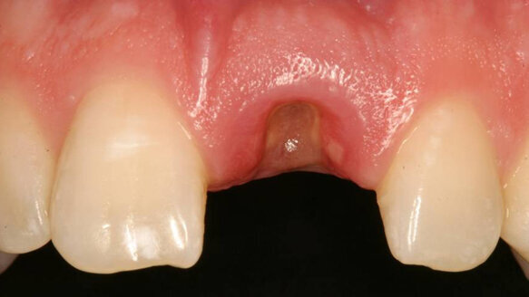
A medically and periodontally stable 37-year-old man presented with coronally fractured tooth #9, which had a history of endodontic treatment (Figs. 1a & b). The tooth was deemed restoratively hopeless.
The treatment plan was as follows:
- Extraction of tooth #9 and socket preservation
- Three-month healing period
- Placement of implant #9 and connective tissue graft
- Three-month healing period
- Implant #9 exposure, placement of healing abutment and connective tissue graft
- Three-month healing period
- Final implant #9 crown restoration
Extraction and socket preservation of tooth #9
After oral sedation with 0.25 mg triazolam one hour prior to surgery and local anesthetic induction using 2 per cent lidocaine with 1:100,000 epinephrine and 0.5 per cent bupivacaine with 1:200,000 epinephrine, a sulcular incision was made circumferentially around tooth #9. The remaining root was extracted atraumatically using a piezoelectric periotome device (Fig. 2).
Thorough degranulation of the extraction site with a pear-shaped carbide finishing bur and Prichard curette proceeded. No dehiscence or fenestration was detected. Freeze-dried bone allograft (FDBA) was used to obliterate the extraction socket.
A bioabsorbable collagen plug (CollaPlug, Zimmer Dental, Carlsbad, CA, USA) was used to cover the graft. The area was secured using 4-0 expanded polytetrafluoroethylene (ePTFE) suture (Fig. 3). The restorative dentist temporized space #9 with an interim removable partial denture.
After three months of uneventful healing (Fig. 4), Stage 1 implant placement was initiated.
#9 fixture placement and connective tissue graft
After oral sedation with 0.25 mg triazolam and local anesthetic induction using 2 per cent lidocaine with 1:100,000 epinephrine and 0.5 per cent bupivacaine with 1:200,000 epinephrine, a flap was created using a trapezoidal papilla-sparing incision design that involved a palatally oriented crestal incision over the #9 site with two vertical releasing incisions made on the buccal, both avoiding the mesial and distal papillae.
A full-thickness flap was raised past the mucogingival junction. Degranulation of the site with a pear-shaped carbide finishing bur and Neumeyer bur revealed adequate apico-coronal, bucco-lingual and mesio-distal dimensions for implant placement.
After osteotomy preparation, a rough-surfaced, internal hex 4 mm (diameter) by 13 mm (length) implant was placed into the filled site (NanoTite Parallel Walled Certain Implant, BIOMET 3i, Palm Beach Gardens, FL, USA) (Fig. 5).
Primary stability was achieved, and a cover screw was placed. In order to form an esthetic soft-tissue profile by expanding mucosal dimensions, a connective tissue graft was harvested from the palate and placed on the buccal aspect of the ridge overlying the implant. The graft was stabilized using 5-0 chromic gut sutures (Fig. 6).
After periosteal release via lateral scalpel incisions, the flap was primarily closed with 4-0 ePTFE sutures in an interrupted and horizontal mattress fashion (Fig. 7). The area was re-temporized with a resin-bonded fixed partial denture.
Implant exposure with connective tissue graft
The #9 site healed well and without incident after three months (Fig. 8). After using a tissue punch technique to remove the mucosa immediately coronal to the fixture (Fig. 9), a one-piece 4.1 mm (platform) by 5 mm (emergence profile) by 4 mm (height) healing abutment (Certain EP Healing Abutment, BIOMET 3i, Palm Beach Gardens, FL, USA) was placed on the #9 implant.
To further augment the buccal ridge dimension, another connective tissue graft was harvested from the palate. A pouch-like envelope flap was raised over the labial ridge aspect into which the connective tissue was transplanted and fixed using 5-0 chromic gut suture (Fig. 10). The healing abutment remained exposed. A periapical radiograph revealed sufficient bone height around the fixture (Fig. 11). The resin-bonded fixed partial denture was replaced.
Final prosthetics
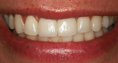
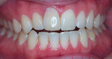
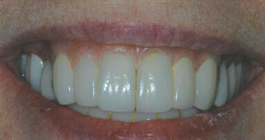
Final restoration of the #9 implant was performed three months post-exposure (Fig. 12). The marginal height and contour of the #9 implant crown matched that of adjacent tooth #8, and a periapical radiograph showed suitable peri-implant bone height (Fig. 13). The patient was satisfied with the functional and esthetic result (Fig. 14).
Postoperative instructions
After each surgical procedure, the patient was instructed to take ibuprofen 600 mg every 4-6 hours, hydrocodone 7.5 mg/acetaminophen 750 mg every 4-6 hours for pain and doxycycline 100 mg qd for 10 days.
The patient was instructed not to brush at or near the surgical site but instead to rinse with 0.12 per cent chlorhexidine or warm saline twice daily. The patient was also directed not to chew in the affected area for at least two weeks. Suture removal occurred at 10 to 14 days post-surgery.
Contact info
Dr Michael Sonick may be contact at +1 203 254 2006 or via his Web site at www.sonickdmd.com.
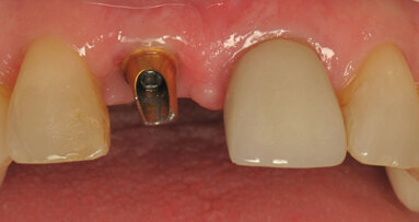
A medically and periodontally stable 24-year-old woman presented with two failing root canals on teeth #8 and #9. Her dental records indicated both teeth ...
Anterior tooth loss and restoration in the esthetic zone is a common challenge in dentistry today. The prominent visibility of the area can be especially ...
NEW YORK, N.Y., USA: In an interview with Endo Tribune U.S. Edition, Emil Jachmann, CEO of Easy-Endo, a materials research company established to capitalize...
When doing a diagnostic work-up, if we line up each challenge that is an obstacle in our quest to provide both a functional and an esthetic end result, each...
In 28 years of placing and restoring implants, I have seen that three key factors need to be present to achieve esthetic implant restorations: good implant ...
Dental fluorosis is an enamel anomaly that adversely affects inorganic phase deposition and organisation, causing enamel hypomineralisation.1 Despite the ...
CHARLOTTE, N.C., US: Dentsply Sirona has announced the US market launch of MIS LYNX, a cost-effective dental implant solution designed by MIS Implants ...
CHICAGO, Ill., USA: As the global leaders in advocating the value and quality of endodontics, the American Association of Endodontists (AAE) supports a ...
ZUG, Switzerland: Late last year, the World Health Organization released a guideline on preventing and managing caries. The document forms part of the ...
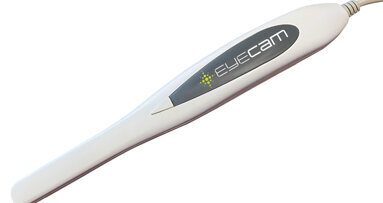
The EyeCam is a fixed focus intraoral camera that produces clear, high-quality images with a single button press, according to Shofu Dental Corp., the ...
Live webinar
Tue. 24 February 2026
1:00 PM EST (New York)
Prof. Dr. Markus B. Hürzeler
Live webinar
Tue. 24 February 2026
3:00 PM EST (New York)
Prof. Dr. Marcel A. Wainwright DDS, PhD
Live webinar
Wed. 25 February 2026
11:00 AM EST (New York)
Prof. Dr. Daniel Edelhoff
Live webinar
Wed. 25 February 2026
1:00 PM EST (New York)
Live webinar
Wed. 25 February 2026
8:00 PM EST (New York)
Live webinar
Tue. 3 March 2026
11:00 AM EST (New York)
Dr. Omar Lugo Cirujano Maxilofacial
Live webinar
Tue. 3 March 2026
8:00 PM EST (New York)
Dr. Vasiliki Maseli DDS, MS, EdM


 Austria / Österreich
 Bosnia and Herzegovina / Босна и Херцеговина
 Bulgaria / България
 Croatia / Hrvatska
 Czech Republic & Slovakia / Česká republika & Slovensko
 France / France
 Germany / Deutschland
 Greece / ΕΛΛΑΔΑ
 Hungary / Hungary
 Italy / Italia
 Netherlands / Nederland
 Nordic / Nordic
 Poland / Polska
 Portugal / Portugal
 Romania & Moldova / România & Moldova
 Slovenia / Slovenija
 Serbia & Montenegro / Србија и Црна Гора
 Spain / España
 Switzerland / Schweiz
 Turkey / Türkiye
 UK & Ireland / UK & Ireland
 International / International
 Brazil / Brasil
 Canada / Canada
 Latin America / Latinoamérica
 China / 中国
 India / भारत गणराज्य
 Pakistan / Pākistān
 Vietnam / Việt Nam
 ASEAN / ASEAN
 Israel / מְדִינַת יִשְׂרָאֵל
 Algeria, Morocco & Tunisia / الجزائر والمغرب وتونس
 Middle East / Middle East












To post a reply please login or register