Search Dental Tribune

Digital workflows for complex adult aligner treatment

Clear aligners and the digital workflow are reshaping adult orthodontics by supporting aesthetic, comfortable and predictable treatment. (Image: EvgeniyQW/AdobeStock; clinical images: Dr Melissa Shotell)

Mon. 1 June 2026

save

Adult orthodontic treatment is considered one of the largest areas of growth in orthodontics. While many orthodontic practices have traditionally been heavily focused on teenage treatment with fixed appliances, some have recognised the need for treatment of adults. As more adult patients are seeking treatment than ever before, the treatment experience of the adult patient has become a new focus in many practices.

Often contrary to their teenage counterparts, adult patients typically seek treatment that is greatly focused on aesthetics and consequently tend to shy away from fixed appliances. Due to their greatly improved aesthetics, clear aligners and transparent brackets have risen in popularity. Recent advances in aligner treatment have addressed the traditional critiques of slower procedures and less anchorage, leading to greater efficiency and comfort during the treatment process.

Further, intra-oral scanning has been widely adopted in orthodontic treatment, not only to aid in the ease of impression procedures, but also to serve as a visual communication tool. Communication with adult patients, especially in setting expectations regarding treatment, has been greatly facilitated by the digital workflow, including intra-oral scanning and visual representation of anticipated outcomes.

Treating adult patients often has unique challenges, including heavily restored dentition, periodontal concerns, medical complexity, long-term parafunctional habits and lifestyle choices such as smoking or consuming alcohol. Many clinicians have turned to aligners as a solution for adult treatment, owing to comfort, ease of hygiene, lack of dietary restrictions on hard and crunchy foods, and communication of expected treatment results. The purpose of this article is to discuss the use of aligner treatment in adult patients and factors that support the use of aligners.

Patient history and clinical findings

A 66-year-old female patient presented for orthodontic consultation as a referral from the prosthodontist treating her. The patient reported past bilateral temporomandibular joint pain associated with bruxism. She had undergone successful splint therapy for temporomandibular disorder (TMD) symptoms and was comfortable with her occlusion, and had a history of lichen planus that was monitored and well controlled. She felt that the crossbite of the mandibular right canine was causing interference with her jaw movement and that her teeth were shifting and the crossbite was becoming worse. The patient expressed a strong interest in aligner treatment so that she would be able to enjoy foods that are to be avoided with fixed appliances and to perform proper oral hygiene. There were also concerns for the potential of mucosal irritation with fixed appliances, and her history of lichen planus.

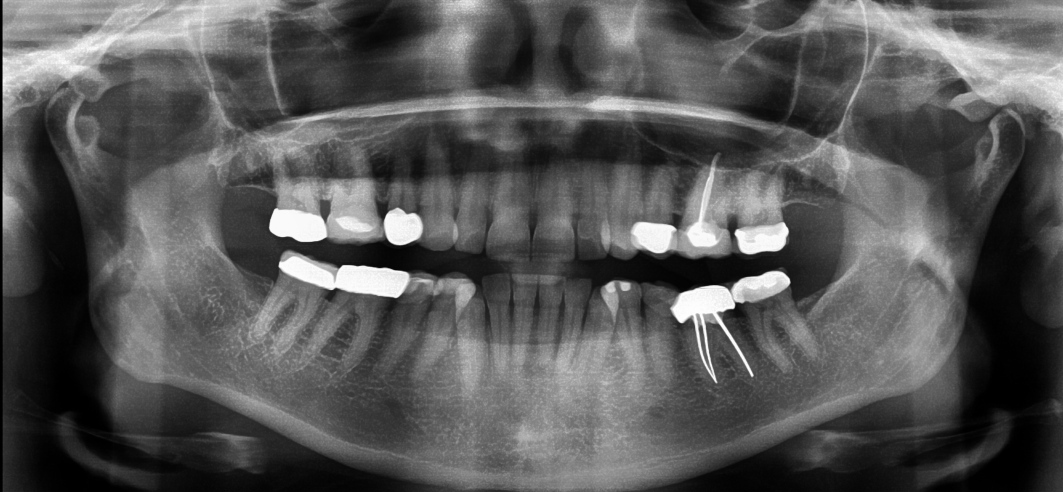
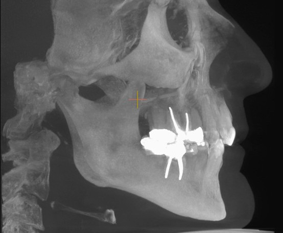
The patient reported orthodontic treatment as a teenager and extraction of two maxillary teeth. Upon examination, both gold and porcelain crowns, several composite restorations and previous extraction of the maxillary first premolars were observed (Figs. 1a–e). She presented with an Angle Class II molar relationship and Class I canine relationship. The mandibular right canine was confirmed to be in crossbite. There was a moderate overbite of 50%, and the mandibular incisors were tipped lingually, creating 4 mm of overjet. The patient appeared to have a relapse of Class II, Division 2 malocclusion in the maxillary arch, evidenced by loss of torque of the central incisors and flaring of the lateral incisors. She had mild crowding in the maxillary arch and moderate crowding in the mandibular arch. The arches lacked symmetry, the maxillary arch being round and the mandibular arch square. The maxillary midline was centred to the face, and the mandibular midline was shifted right by 3 mm. There was no detectable shift between centric occlusion and centric relation, and the patient had been restored to a cusp-to-fossa functional relationship. The patient appeared to have a restricted envelope of function due to the crossbite of the mandibular right canine and the Class II, Division 2 relapse of the maxillary malocclusion.

Fig. 2a: Pretreatment panoramic radiograph.

Fig. 2b: Pretreatment lateral cephalogram.

Radiographic examination with a panoramic radiograph reconstructed from CBCT data and a traditional lateral cephalogram showed teeth restored with crowns and endodontically treated teeth in the posterior, as well as regular root morphology (Figs. 2a & b). The panoramic radiograph showed a fully erupted adult dentition from second molar to second molar, and the lateral cephalogram showed a Class I skeletal relationship. The temporomandibular joints were well corticated on radiographic examination, and there was excess bone below the head of the condyles, as expected considering the history of parafunction. There were no signs of temporomandibular joint pathology. The patient was periodontally healthy and demonstrated excellent oral hygiene for orthodontic treatment.

Treatment options: Fixed appliances versus aligners

After discussion of the findings of the clinical examination, the patient was presented with two treatment options. The first would have involved the extraction of the mandibular right central incisor and would have resolved the crossbite utilising the extraction space. Further, the mandibular midline would have been centred on the mandibular left central incisor. The second option would have utilised interproximal reduction (IPR) from canine to canine and proclination of the mandibular incisors, but the mandibular midline would not have been coincident with the maxillary midline.

The patient was also given the option of traditional bracket and wire treatment or aligner treatment using ClearCorrect (Straumann). In the treatment discussion, the patient expressed her concern about her previous TMD symptoms. She felt that her posterior occlusion was comfortable after splint therapy, and she did not want to have her posterior restorations replaced. The patient also expressed concerns of possible mucosal irritation with fixed appliances, particularly in light of her history of lichen planus. For these reasons, we felt that aligner treatment would be advantageous for this patient, and the flexibility of being able to remove the aligners during any acute TMD flare-up during treatment was a strong deciding factor.

The patient was not concerned about addressing the mandibular midline and was primarily looking for an aesthetically pleasing smile and resolution of the crossbite of the mandibular right canine. The focus of treatment would be to resolve the mandibular right canine crossbite. We reviewed the patient’s limited goals for treatment and her desire to complete treatment quickly and determined that the patient would be most comfortable and well served with nonextraction aligner treatment limited to the anterior teeth. She agreed that final retention would utilise a maxillary splint if her TMD symptoms returned.

A treatment plan with the following treatment goals was established:

  1. resolve the crossbite of the mandibular right canine;
  2. maintain the posterior restored occlusion;
  3. resolve the crowding;
  4. improve the excess overbite;
  5. improve the overjet;
  6. improve the mandibular midline;
  7. improve the smile aesthetics; and
  8. improve the maxillary incisor torque to improve the envelope of function.

A digital treatment planning approach was utilised, including the taking of diagnostic impressions with a TRIOS intra-oral scanner (3Shape; Fig. 3), photographs using an EOS Rebel T6i DSLR camera (Canon) and a 15 × 15 cm CBCT scan using a Green CT2 scanner (Vatech). Use of intra-oral scanning gave greater patient comfort and allowed for a faster impression procedure to avoid the amount of time the patient would have her mouth open and to thus reduce the risk of potentially exacerbating her previous TMD symptoms. The combination of intra-oral and CBCT scans and photographs supports patient communication and helps to explain the current state of the malocclusion and discuss goals for treatment.

Fig. 3: Intra-oral scan taken with the TRIOS (3Shape).

Fig. 3: Intra-oral scan taken with the TRIOS (3Shape).

Fig. 4: Initial ClearCorrect treatment set-up. Anticipated treatment results after 12 steps of aligner treatment.

Fig. 4: Initial ClearCorrect treatment set-up. Anticipated treatment results after 12 steps of aligner treatment.

Fig. 5a: Interproximal reduction and attachments planned with ClearCorrect aligners, frontal view.

Fig. 5a: Interproximal reduction and attachments planned with ClearCorrect aligners, frontal view.

The aligner system

Fig. 5b: Interproximal reduction and attachments planned with ClearCorrect aligners, occlusal view.

The ClearCorrect system was selected for this patient based upon several clinical considerations. Based on the patient’s history of lichen planus, we wanted to limit the use of attachments (engagers) and time in treatment and to avoid bonding attachments to the porcelain and gold restorations. The straight extended trim line gives the aligners increased retention while accomplishing the aforementioned reduction of attachments.1–3 The threelayer material, ClearQuartz, also gives the aligner a lower insertion force and greater sustained force delivery.4–7 This creates a system with increased comfort and increased activation time per aligner. The use of the aligner system allowed us to reduce the number of attachments and treatment steps and to provide a course of treatment with enhanced patient comfort and was supported by the digital workflow.

Digital workflow and treatment

The intra-oral scans and photographs were submitted to ClearCorrect to create a treatment set-up. The first set-up involved 12 treatment steps and utilised attachments on the maxillary and mandibular anterior teeth to aid intrusive and rotational movements (Fig. 4). Interproximal reduction (IPR) of 1.4 mm in total, was planned from the distal aspect of the mandibular left canine to the distal aspect of the right canine to facilitate resolution of the crowding and correction of the crossbite of the mandibular right canine (Figs. 5a & b). The mandibular incisors were also proclined in order to aid in resolution of the crowding and to improve the overbite and overjet. Aligners were planned to be worn for 22 hours per day, and each aligner was to be worn for a 14-day wear cycle.

The aligners were delivered to the patient with instructions for home care and daily wear. The patient was also given elastomeric aligner seating aids, aligner chews, but was instructed to discontinue their use if any symptoms of TMD were experienced. The patient was given the first two sets of aligners and rescheduled for bonding of attachments four weeks later. The attachments were bonded using Assure Plus bonding agent and GoTo bracket paste (Reliance Orthodontic Products). The patient was seen for periodic aligner checks, and the IPR was performed in two phases at the seventh and ninth treatment steps, to avoid discomfort and prolonged mouth opening.

The patient completed the initial 12 steps of aligner treatment in six months. The aligners fitted well at completion of the first phase of aligner treatment. At the progress evaluation visit, it was determined that additional improvement in tooth position could be achieved, including additional correction of the mandibular midline, improvement of the mandibular right canine position and improvement in arch coordination. The attachments were removed, and progress records were taken, including photographs (Figs. 6a–e) and intra-oral scans. The progress records were sent to ClearCorrect, and treatment revision was requested. Three additional aligners were planned, and an attachment on the mandibular right canine to aid extrusion for improved canine guidance (Fig. 7). Detailing of the incisor position was planned for smile aesthetics, and no additional IPR was utilised.

The patient completed her revision aligner treatment in six weeks and was not seen during this time. An office visit was scheduled for the completion of the revision aligner sequence, and at this visit, the attachments were removed, and the patient was placed into final retention with retainers made by ClearCorrect.

Treatment results

Treatment was completed in nine months using 15 sets of aligners (Figs. 8a–e). The final treatment results addressed the patient’s chief complaint and fulfilled her goals and expectations. Arch symmetry was improved, the crowding and excess overbite were resolved.

The excess overjet and the crossbite of the mandibular right canine were improved (Figs. 9a–f). The patient reported that her TMD symptoms remained stable and that she was comfortable in aligner retention. The patient indicated that her aesthetics were greatly improved and that she was incredibly pleased with her overall treatment results and her aligner treatment experience.

Figs. 9a–f: Photographs before and after aligner treatment showing resolved crowding and crossbite of the mandibular right canine, as well as improved overbite, overjet and arch symmetry. Before (a).

Figs. 9a–f: Photographs before and after aligner treatment showing resolved crowding and crossbite of the mandibular right canine, as well as improved overbite, overjet and arch symmetry. Before (a).

Fig. 9b: After.

Fig. 9b: After.

Fig. 9c: Before.

Fig. 9c: Before.

Fig. 9d: After.

Fig. 9d: After.

Fig. 9e: Before.

Fig. 9e: Before.

Fig. 9f: After.

Fig. 9f: After.

Conclusion

ClearCorrect aligners were able to deliver the treatment results that the patient was expecting and in a limited amount of time. The use of a unique aligner design and materials allowed us to limit the number of treatment steps and attachments used. Further, selective areas of IPR and near-continuous wear allowed for resolution of the crowding and of the crossbite of the mandibular right canine while improving canine-guided occlusion. The aligner treatment resulted in an improvement in arch symmetry, occlusion, the envelope of function and smile aesthetics. The high level of patient compliance, combined with the design and materials of the ClearCorrect system, allowed the patient to complete treatment in a short period and to have excellent results and experience.

Editorial note:

The list of references can be found here. This article was published in aligners—international magazine of aligner orthodontics vol. 5, issue 1/2026.

Topics:
Tags:
To post a reply please login or register
advertisement
advertisement